ADENOIOD
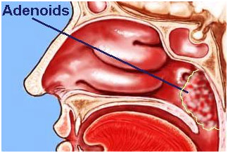
Adenoids are lymphoid follicular collection situated in the nasopharynx (space at the back of the nose). Usually they regress and do not cause any problem. Occasionally the size of the adenoid may be large enough which leads to nasal blockage, mouth breathing and due to this frequent infections of the throat and ear involvement may occur
Effects on ears:
The enlarged adenoid may lead to hearing impairment, pain in the ears, and discharge if not treated properly. This happens due to the malfunction of the eustachiantubes (the tube which connects the middle ear to the back of the nose). The tube usually allow the air to come out and get in the of the middle ear so to maintain normal pressure in the middle ear. This is noticed while landingof anaero plane. The constant negative pressure in the middle ear as a result of the enlarged adenoids hinders the function of the eustachian tubes, the watery part of the blood (plasma) accumulates in the middle ear. This fluid in the middle ear limits the free movement of the ossicles i.e the chain of bones connected to the ear drum and with the movement of this chain of bones the fluid in the inner ear (cochlea ) moves and an electric impulse is generated which is sent to the brain via the auditory nerve. Through the nerve impulse reaches the brain for full differentiation. Sometimes bacteria get into the fluid accumulated in the middle ear and this results in severe pain and the ear drum stretches and the bulge may cause rupture of the ear drum(tympanic membrane).The rupture of tympanic membrane reduces the pain but the discharge occurs which may persists and a perforation is developed which may require surgical repair by plastic surgery known as tympanoplasty
If the infection doesnot occur and if the fluid is accumulated in the middle ear stays for a longer time, it becomes more viscous and the condition is called Glue ears. With the formation of glue the movements of the ossicles (chain bones connected to the tympanic membrane) is reduced.
Diagnosis of Glue ears:
The deafness caused by the glue ears is diagnosed by clinical examination and investigations include Tympanometry or the impedence tests.
Treatment of Glue ears:
Treatment of the Glue ear is to remove the glue and insert a ventilating tube which is called grommet. This procedure is performed under general anesthesia. First the ear drum is incised (this part of the procedure is called Myringotomy). After the incision of the ear drum the glue is sucked out and the grommet ( ventilation tube) is inserted. See the sketch below:
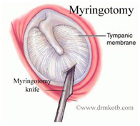
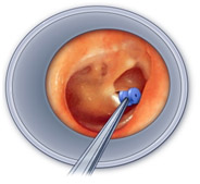
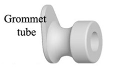
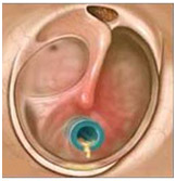
Ear drums is incised under general anesthesia and a ventilating tube is inserted in the ear drum. This tube remains in the ear for 3 months to more than one year depending on the shape of the ventilating tube.
Sometimes the glue ears occur with enlarged adenoids and one has to perform the grommets surgery for ventilation of the middle ear. The glue ears if not treated properly will lead to a condition known as chromic adhesive otitis media, where the tympanic membrane becomes adherent to the promontory and the deafness becomes permanent.
Bad effects airway:
Enlarged adenoids cause air way obstruction and this is noticed as restless at night and during sleep noises and snoring occurs. The sleep apnea is of concern i.e. there is breath holding and this cessationstoppage of breathing during sleep for more than seven seconds (known as apnea) has ill effects on the health and mind. Apnea may lead to increase in blood pressure and lack of concentration on and memory loss.
The other known effect of mouth breathing is the frequent tonsillitis and upper air way infections.
Surgery of Adenoids:
The adenoids surgery is done under general anaesthesia. The surgeon sits at the head end of the patient and after opening the mouth with a mouth gag the adenoid currete is applied to take out the adenoids. This is the conventional surgery still done at many centers. Here the surgeon has no direct visions of the adenoids and the bleeding used to be stopped by putting a pack and repeated packs till it stops or otherwise a packing has to be left in foe 24 to 48 hours.
The conventional adenoidectomy bears the risk of injury to Eustachian tube or posterior choanae since the procedure is either blind or by indirect visualization. The removal at times remains incomplete. The use of endoscope and laser makes the procedure more effective and safe.
Adenoidectomy can be performed with variety of instruments. All these methods have their advantages and disadvantages. I believe the choice of instruments is usually based solely on the surgeons’ personal preference and availability.
My techniqueI perform adenoid surgery under the camera vision and I use Diode Laser at the start of surgery. Diode laser has the advantage that it is delivered on a fiber. I expose the bed of the adenoid to Diode laser under vision of the endoscope . I curate the adenoids and the curette is done once only. Any remaining remnants of adenoids are not curetted again as multiple curettage leads to post operative pain. Instead, I use the Micro debrizor which is also called shaver to pick the remaining adenoid tissue. This motorized instrument is very helpful in the complete removal of the adenoid tissue. Finally the oozing from adenoid bed is subjected to Diode Laser again to achieve complete hemostasis.
The benefit of my technique is definitely there with minimum blood loss, less pain and complete removal of the disease and complete irradiation recurrence of the disease and minimal recurrence. The disadvantage is the cost of the disposable blades of the micro debrizor and the costly laser machine.
I must ask myself whether a more precise, more complete and more satisfying resection with less blood loss, early recovery and less pain justifies the additional cost or not.